Pelvic Anatomy - "Anatomy of human pelvic bone." Art Print by ...
Pelvic Anatomy - "Anatomy of human pelvic bone." Art Print by .... Because of the pelvis' important role in obstetrics, it is one of the most sexually dimorphic bony elements of the human body. There are four articulations within the pelvis: Wasnik, mbbs, mda, michael b. It is further divided into the greater (false) and lesser (true) pelvis. The pelvic region is the area between the trunk — or main body — and the lower extremities, or legs.
ads/bitcoin1.txt
• divided into the true and false pelvis by the iliopectineal line. The pelvic bones are smaller and narrower. For an average risk woman, this is offered starting at age 21, and is repeated every 3 years if the findings are normal. The pelvis is an anatomically complex and functionally informative bone that contributes directly to both human locomotion and obstetrics. Until puberty, each hip bone consists of three separate bones yet to be fused:
Pelvic Girdle (With images) | Gross anatomy, Pelvic girdle ... from i.pinimg.com The pelvic region is the area between the trunk — or main body — and the lower extremities, or legs. The bony pelvis is formed by the sacrum and coccyx and a pair of hip bones (ossa coxae), which are part of the appendicular skeleton.its primary function is the transmission of forces from the axial skeleton to the lower limbs as well as supporting the pelvic viscera. However, knowledge of the anatomy of various structures that surround these organs has evolved over time. • divided into the true and false pelvis by the iliopectineal line. This mri male pelvis axial cross sectional anatomy tool is absolutely free to use. The pelvic exam is typically done in a number of settings: Two female reproductive organs located in the pelvis. Learn from international mis surgeons with didactic anatomy dissection on female cadavers and gain hands on experience.
The male pelvis is different from a female's.
ads/bitcoin2.txt
It is strengthened and supported by several joints and ligaments. The lumbosacral plexus is formed by the lumbosacral trunk and the ventral rami of the first to third sacral nerves, and part of the fourth sacral nerve. Although ultrasound is frequently indicated for the primary evaluation of The male urethra and the penis The pelvis is a basin shaped bony structure formed by the combination of two pelvic bones (hip bones or innominate bones) and the sacrum. It also extends the leg (and/or thigh) at the knee joint. • pelvis begins at the iliac crests and ends at the symphysis pubis. The pelvic girdle and pelvic spine. The lining of the uterus. Pelvic anatomy on mri ashish p. Two female reproductive organs located in the pelvis. • located inferior to the pelvic brim. A pelvis mri (magnetic resonance imaging) scan is an imaging test that uses a machine with powerful magnets and radio waves to create pictures of the area between the hip bones.
As part of cervical cancer screening, which involves a pap smear. Because of the pelvis' important role in obstetrics, it is one of the most sexually dimorphic bony elements of the human body. The lining of the uterus. True pelvis • also known as pelvic cavity. The pelvis is the lower portion of the trunk, located between the abdomen and the lower limbs.
Pelvic Floor Anatomy - Pelvic Diaphragm part 1 - YouTube from i.ytimg.com The pelvis (plural pelves or pelvises) is either the lower part of the trunk of the human body between the abdomen and the thighs (sometimes also called pelvic region of the trunk) or the skeleton embedded in it (sometimes also called bony pelvis, or pelvic skeleton). • located inferior to the pelvic brim. Because of the pelvis' important role in obstetrics, it is one of the most sexually dimorphic bony elements of the human body. The first step is assessing the mass' site of origin and its location in relation to the peritoneal cavity and the extraperitoneal spaces. The male pelvis is different from a female's. The male pelvic floor is a complex structure made up of muscles, ligaments, nerves and fascia. A pelvic ultrasound is a noninvasive diagnostic exam that produces images that are used to assess organs and structures within the female pelvis. The pelvic girdle and pelvic spine.
• pelvis begins at the iliac crests and ends at the symphysis pubis.
ads/bitcoin2.txt
The testicles and scrotum are also important male structures. For an average risk woman, this is offered starting at age 21, and is repeated every 3 years if the findings are normal. The rectus femoris flexes the thigh at the hip joint and anteriorly tilts the pelvis at the hip joint. This area provides support for the intestines and also contains the bladder and reproductive organs. Until puberty, each hip bone consists of three separate bones yet to be fused: True pelvis • also known as pelvic cavity. The right and left hip bones also converge anteriorly to attach to each other. Although ultrasound is frequently indicated for the primary evaluation of The anatomy of the pelvis varies depending on whether you are male or female. Anatomy of female pelvic area. The pelvis is an anatomically complex and functionally informative bone that contributes directly to both human locomotion and obstetrics. The lining of the uterus. The male urethra and the penis
Imaios and selected third parties, use cookies or similar technologies, in particular for audience measurement. The male pelvis is different from a female's. The pelvic region is the area between the trunk — or main body — and the lower extremities, or legs. True pelvis • also known as pelvic cavity. The bony pelvis consists of the two hip bones (also known as innominate or pelvic bones), the sacrum and the coccyx.
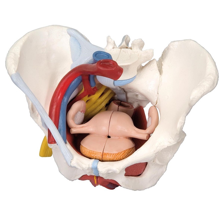
Anatomical Models of Female Pelvis with Ligaments, Vessels ... from www.mentone-educational.com.au This mri male pelvis axial cross sectional anatomy tool is absolutely free to use. It is usually divided into two separate anatomic regions: Wasnik, mbbs, mda, michael b. The bony pelvis consists of the two hip bones (also known as innominate or pelvic bones), the sacrum and the coccyx. Pelvic anatomy on mri ashish p. The rectus femoris flexes the thigh at the hip joint and anteriorly tilts the pelvis at the hip joint. A pelvic ultrasound allows quick visualization of the female pelvic organs and structures including the uterus, cervix, vagina, fallopian tubes and ovaries. As part of cervical cancer screening, which involves a pap smear.
The pelvis (plural pelves or pelvises) is either the lower part of the trunk of the human body between the abdomen and the thighs (sometimes also called pelvic region of the trunk) or the skeleton embedded in it (sometimes also called bony pelvis, or pelvic skeleton).
ads/bitcoin2.txt
Two female reproductive organs located in the pelvis. Visualise your pelvic floor and see exactly what it is, where it's located and why it is important to train this hidden group of muscles. The bony pelvis consists of the two hip bones (also known as innominate or pelvic bones), the sacrum and the coccyx. It is strengthened and supported by several joints and ligaments. • located inferior to the pelvic brim. This part of the body is called the pelvic area. The pelvic girdle (hip girdle) is formed by a single bone, the hip bone or coxal bone (coxal = hip), which serves as the attachment point for each lower limb. Pelvic anatomy on mri ashish p. However, knowledge of the anatomy of various structures that surround these organs has evolved over time. The male urethra and the penis • divided into the true and false pelvis by the iliopectineal line. This area provides support for the intestines and also contains the bladder and reproductive organs. Structures inside and near the pelvis include the bladder, prostate and other male reproductive organs.
ads/bitcoin3.txt
ads/bitcoin4.txt
ads/bitcoin5.txt
0 Response to "Pelvic Anatomy - "Anatomy of human pelvic bone." Art Print by ..."


0 Response to "Pelvic Anatomy - "Anatomy of human pelvic bone." Art Print by ..."
Post a Comment